Severe digital gangrene – when to include tromboangiitis obliterans on the diagnosis list?
Image Description
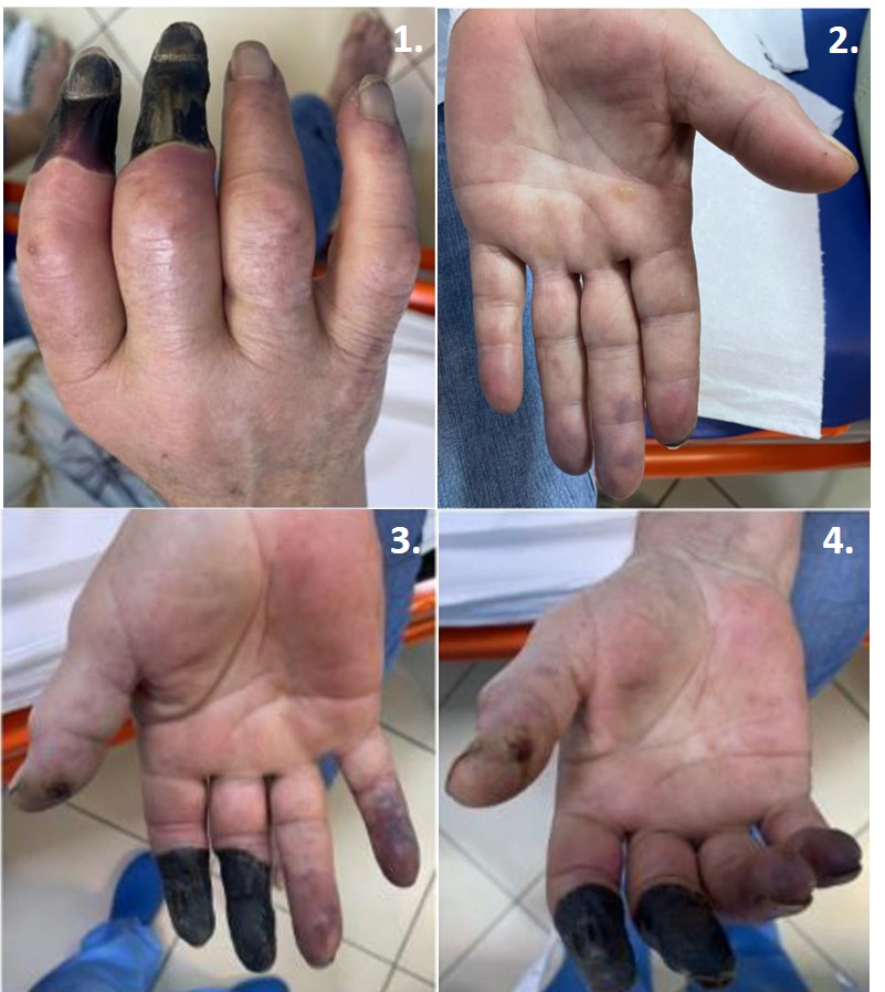
A 63-year-old female patient presented to the Emergency Unit for severe necrosis of two digits in right hand and signs of ischemia in the remaining digits, left hand and feet. She complained of inflammatory-like arthralgias in her wrists and claudication while walking but denied history of Raynaud’s phenomenon. Apart from having a well-controlled arterial hypertension and type 2 diabetes, patient was a heavy smoker (approximately 80 pack-year). First skin color changes suggesting cyanosis in peripheral upper limbs occurred three weeks prior, accompanied by pain, local low temperature and diminished pulse. Laboratory markers showed high inflammatory marker values and moderate leukocytosis, while immunological investigations ruled out the presence of antinuclear antibodies (ANA), antineutrophil cytoplasmic antibodies (ANCA), cryoglobulins and anti-phospholipid antibodies. Heart ultrasound showed no signs of endocarditis. The Doppler ultrasound confirmed normal flow velocities with only mild, non-impairing atheromatosis. Capillaroscopy did not identify specific features.
Patient was referred to a surgical department for intravenous prostaglandins and local management of the necrotic areas.
The diagnosis of tromboangiitis obliterans (TAO) was considered. TAO or Buerger’s disease is a rare vasculitis that affects small and medium-size arteries and veins of the upper and lower limbs and occurs in young heavy smokers (1). Diagnosis is based on clinical changes and confirmed through arteriography that shows segmental involvement of vessels and corkscrew-like vascularization around the area of occlusion (2). Treatment encompasses smoking cessation, vasodilators like prostanoids or phosphodiesterase inhibitors, while revascularization has not yet proved as beneficial (3).
References
Tanaka K. Pathology and pathogenesis of Buerger’s disease. Int J Cardiol. 1998 Oct 1;66(SUPPL. 1).
Mollet NR, Cademartiri F, Van Mieghem CAG, Runza G, McFadden EP, Baks T, et al. High-resolution spiral computed tomography coronary angiography in patients referred for diagnostic conventional coronary angiography. Circulation. 2005 Oct 11;112(15):2318–23.
Mitropoulos F, Eforakopoulos F, Kanakis MA, Vassili M, Mastorakou I, Georgiadis M. Diagnostic and therapeutic approach in a patient with Buerger’s and coronary artery disease. Case Rep Med. 2013;2013.