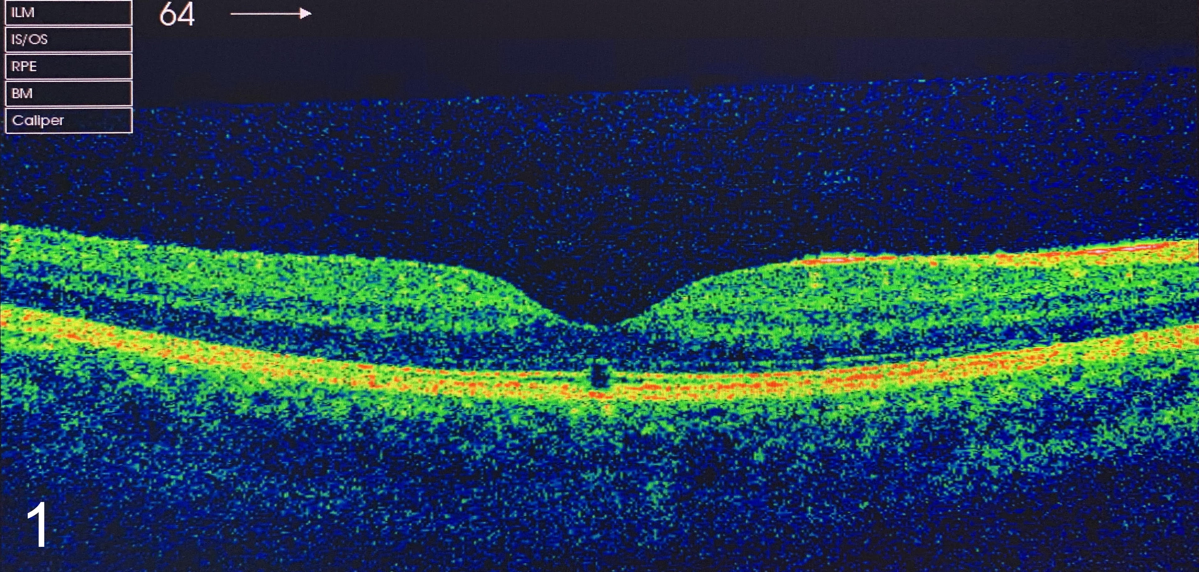
Optical coherence tomography showing retinal damage from handheld green diode laser pointer
Image Description
An 8-year-old boy was brought with impaired vision in the right eye, and a paracentral scotoma after being accidentally exposed to a class IIIA green diode laser pointer, of 650 nm wavelength and 5 mW power, and directly viewed the green light for several seconds, two days before presentation. His best-corrected visual acuity in both eyes was 6/7.5 on the Snellen chart. Slit-lamp examination revealed a normal anterior pole in both eyes, and fundoscopy, was normal as well. Macular OCT (optical coherence tomography) scan revealed focal interruption involving the photoreceptor inner segment - outer segment junction, extended toward the retinal pigment epithelium.
Green diode lasers can produce permanent visual loss due to retinal damage, and should not be used by children. The ophthalmic community, as well as the public, should be aware of the potential danger of this handheld laser if it is improperly used. (1)
References
(1) Sell CH, Bryan JS. Maculopathy From Handheld Diode Laser Pointer. Arch Ophthalmol. 1999;117(11):1557–1558.