Appearances may deceive: diagnosing a polypoid adenomyoma of the uterus
Image Description
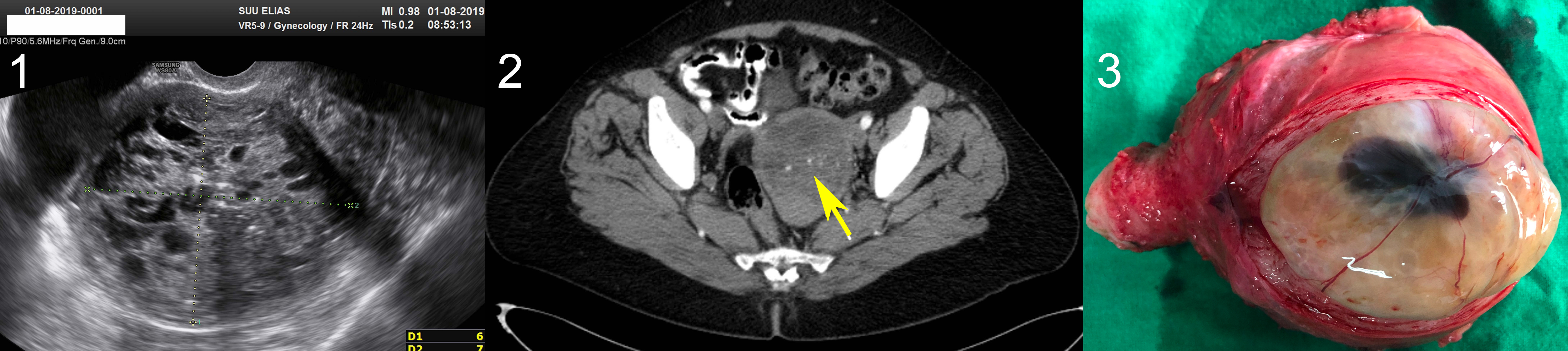
This is the case of a 68-year old menopausal female who accused abdominal pain and transient vaginal bleeding over the past three months. Vaginal ultrasound showed an enlarged uterus, with thin walls and a heterogeneous mass in the cavity (Fig. 1), with visible blood flow on Doppler exam, suggesting endometrial cancer. The computed tomography confirmed a large mass developed in the uterus, with benign features, suggesting uterine myoma (Fig. 2). The endometrial biopsy showed an endometrial surface epithelium, without nuclear atypia and fragments of immature squamous metaplasia epithelium, without cytonuclear atypia. Laparotomic hysterectomy was performed. The postoperative macroscopic exam of the uterus after longitudinal section showed a polycystic tumour of 5/4/4 cm (Fig. 3). The postoperative pathological report found an endometrial polyp with loose areas, edematous, with cystic dilated glands and mature and focal immature metaplasia in the epithelium and some glands, consistent with a polypoid adenomyoma. The sonographic feature in our case suggested endometrial cancer, due to the poorly defined margin with the underlying myometrium, the presence of multiple small cysts and a heterogeneous echotexture. Still, uterine myomas frequently undergo degenerative changes, which significantly alter their imaging appearances, thus uncommon modifications in menopausal women may be difficult to differentiate from malignant endometrial tumours, based solely on imaging and clinical feature. Polypoid adenomyomas aren’t always easy to differentiate from other tumours based on sonography, thus familiarity with these characteristics must draw attention in the process of reaching the correct diagnosis.