Intralenticular foreign body
Image Description
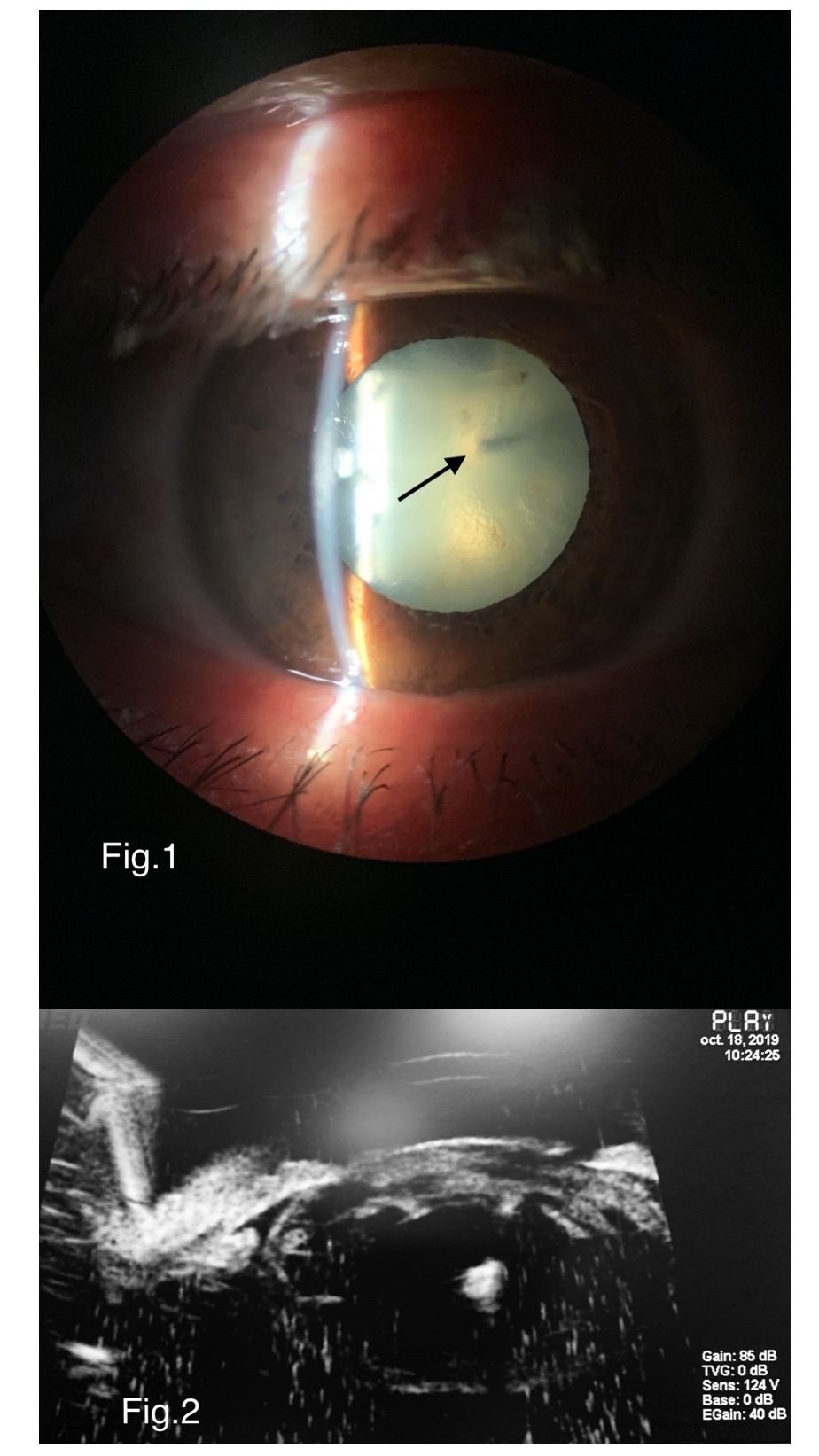
A 46 years old man presented with impaired vision in the right eye for about 6 months, chemosis and hyperlacrimation for 2 months. His best-corrected visual acuity was 6/75 on Snellen chart. Anterior pole slit-lamp examination revealed a small self-sealed corneal wound at the 1 o’clock position, corticonuclear opacification of the intraocular lens, with anterior capsule tear also at the 1 o’clock position, and a paracentral metallic foreign body in the upper part of the nucleus (Fig.1). Intraocular pressure was normal. Fundoscopy of the right eye showed a normal eye fundus. Cranial CT scan revealed no other associated abnormalities. The Ultrasound Biomicroscopy image showed a highly echogenic intralenticular mass (Fig.2). Anterior segment and eye fundus of the left eye were normal. Despite anterior capture rupture, the rhexis was successfully made, cataract surgery and removal of the foreign body were performed, with posterior chamber intraocular lens implantation. The visual outcome was good, with a 6/6 best-corrected visual acuity on the Snellen chart. Local antibiotics and steroids were administered.
Intralenticular foreign body is rarely seen in practice. Conservative management is a good option unless the visual function is compromised (1). The most serious complication of a retained metallic foreign body containing iron, is siderosis oculi, with a very poor visual prognosis (2).
References
(1) Bowling B, Kanski’s Clinical Ophtalmology, 2016, ELSEVIER, p 881
(2) Reddy SC. Intralenticular metallic foreign body: a case report. Int J Ophthalmol. 2011;4(3):326–328. doi:10.3980/j.issn.2222-3959.2011.03.25