Left hepatic lobe necrosis after laparoscopic treatment of a hiatal hernia
Image Description
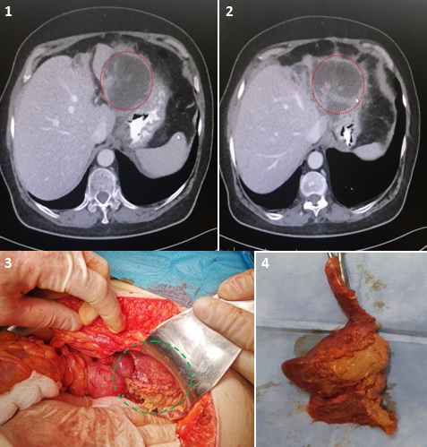
The patient aged 66 years was admitted to the General Surgery Clinic for pain in the upper abdominal quadrant with posterior irradiation and weight loss. Two and a half years before the patient underwent surgery for a hiatal hernia when a Nissen fundoplication was performed by laparoscopic approach. CT was performed which confirmed the presence of a large abscess at the level of the left hepatic lobe and lobe atrophy (Figure 1, Figure 2). Surgery was undertaken. Abdominal exploration identified ischemia and necrosis of segments 2 and 3 of the liver so a resection was decided upon (Figure 3, Figure 4). The postoperative evolution was favourable. NB: Ischemic necrosis of the left hepatic lobe most likely occurred after the surgical treatment of the hiatal hernia when the left hepatic artery was resected on dissection which was probably the only source of blood supply for the left lobe. It is important that these vascular abnormalities are known by the surgeon so during dissection of the gastrohepatic ligament any arteries identified should be protected to avoid possible necrosis of the left hepatic lobe.