Inside of an adrenal cyst
Image Description
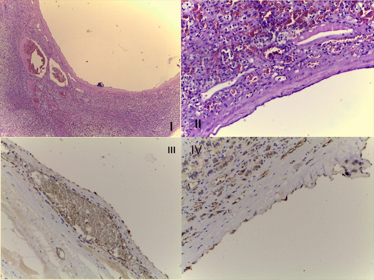
This is a 36-year old menstruating female who accused abdominal pain of non-specific pattern during the last days. Abdominal ultrasound and computed tomography confirmed a left adrenal mass of 6 centimetres with cystic appearance without solid elements. No other causes of abdominal pain were identified. Adrenalectomy was performed. The pre- and post-operatory panel of endocrine assessments were normal. The pathological report confirmed an endothelial cyst at the level of a normal adrenal gland. Hematoxylin-eosin staining is used for the epithelial cells (Figure I: 4X, Figure II: 40X). The immunochemistry reaction is positive for factor VIII in cyst cells of adrenal origin. (Figure III: FVIII positive immunostain in fusiform and cubic cells, 20X, Figure IV: FVIII in fusiform cells, 20X). Further on the clinical evolution was good. Adrenal cysts may be endothelial related, epithelial-derived and pseudocysts. They are found in both adults and children but the level of evidence varies from cases series to cases reports. (1,2,3) The risk of malignancy or rupture is a major complication. (1) Endothelial cysts seem more frequent in women. (2) The scenario of detection may be of a typical adrenal incidentaloma or starting from non-specific local as seen here.