Superficial Spreading Melanoma: Clinical Case, Histological Analysis, and Management
Image Description
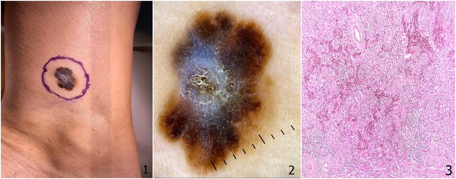
Skin cancer, particularly melanoma, is a serious health concern that demands timely diagnosis and treatment. We present the case of a 36-year-old woman who carries a family medical history of cancer. Her journey to diagnosis commenced with a routine dermatoscopic examination, revealing an atypical nevus in an unexpected location - a solitary tumor, approximately 1.5 cm in diameter, in the left internal supramalleolar region (Fig.1), that exhibited several distinctive dermoscopic features, including irregular margins, chromatic polymorphism, an atypical pigment network, blue-white veil with a central regression area, uneven dots and globules (Fig. 2). Recognizing the need for thorough evaluation, a surgical excision was promptly performed, maintaining a 0.5 cm safety margin. The histopathological examination (Fig. 3) unveiled primary cutaneous melanoma, classified as unulcerated, with a Breslow thickness of 1.2 mm (pT2a) and Clark Level III invasion. The histological subtype revealed superficial spreading melanoma, with a low dermic mitotic index (1 mitosis per square millimeter), with no lymphovascular invasion or neurotropism. The melanoma exhibited partial regression. Immunohistochemistry revealed Melan A's diffuse positivity, SOX10's nuclear-positive tumor cells without emboli or microsatellites, PRAME's nuclear-positivity, and p16's abnormal expression in tumor cells but positivity in the hyperplastic epidermis.
This case highlights melanoma's diverse characteristics, demonstrating its ability to appear in unexpected areas with distinctive clinical and pathological attributes. Regular dermatoscopic monitoring is a valuable tool for tracking the patient's progress and detecting any potential issues at an early stage. Histological analysis and immunohistochemistry are crucial for precise diagnosis and should not be undervalued.
References
Trindade FM, de Freitas MLP, Bittencourt FV. Dermoscopic evaluation of superficial spreading melanoma. An Bras Dermatol. 2021;96(2):139–47. doi:10.1016/j.abd.2020.06.012 Journal
Melanoma Treatment (PDQ®)–Health Professional Version. National Cancer Institute. Available at https://www.cancer.gov/types/skin/hp/melanoma-treatment-pdq#link/_25_toc. October 14, 2022; Accessed: January 26, 2023.
Wilson ML. Histopathologic and Molecular Diagnosis of Melanoma. Clin Plast Surg. 2021 Oct. 48 (4):587-598. [QxMD MEDLINE Link].